Tuesday, May 22, 2018

Armpit Rolls and Tail of the Breast

Please tweet and retweet


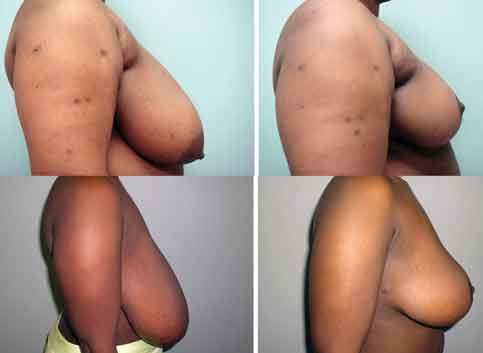
Armpit Rolls
Armpit rolls are folds of skin and fat that lie at the upper front edge of the armpit. They make some women self conscious about wearing spaghetti strap tops or strapless dresses and bras. Their presence can be independent of breast size or patient weight
armpit rolls are independent of breast size
Armpit Rolls Are Independent of Breast Size as Seen Before and After Breast Reduction Surgery


Wednesday, April 4, 2018

Seborrheic Keratosis/Keratoses - Warts

Please tweet and retweet


Seborrheic keratoses are common superficial skin tumors made up of immature superficial skin cells and sometimes referred to as warts. They usually develop after the age of 50, but they can also appear in young adulthood. There is a genetic predisposition to develop a high number of seborrheic keratoses, although the precise inheritance pattern is unknown. Their cause is unknown but UV radiation/sun exposure or human papillomavirus (HPV) infection are suspected. They have well defined borders and are well-demarcated, round or oval skin lesions with a dull, uneven cauliflower like surface and a typical stuck-on appearance. Over time they can grow and become darker in color. Virtually everyone will have these if they live long enough.

They generally do not cause any symptoms, but chronic irritation due to friction trauma may occasionally cause itching, pain, or bleeding. The diagnosis of seborrheic keratosis is usually based on their appearance of being "stuck on," warty, distinctly margined, often scaly dark superficial skin lesions located most commonly on the trunk, face, and upper extremities but they can occur anywhere that you have skin including the scalp. Since they are benign and slow growing health insurance does not cover their removal unless there is a suspicion of skin cancer or the keratosis becomes infected etc. While most are clearly visible as keratoses some keratoses are hard to differentiate from a cancer in which case a biopsy is required to know for sure.

The most common treatment is freezing (cryotherapy) followed by shaving, excision, electrodessication (burning with an electric current) or burning it off with a laser. Other than shaving or excision the other methods of treatment destroy the tissue so a definitive diagnosis under the microscope is not possible. Also each of these methods of removal leave a scar. Cryotherapy also removes skin pigment leaving you with a visible white spot so its use is limited in darker skinned individuals.

A new treatment has recently been FDA approved and is now available in the US.
Eskata is a 40% hydrogen peroxide solution dispensed in a pen brush applicator. 2 applications (4 times each 1 minute apart per visit) 2 to 3 weeks apart completely removes most seborrheic keratoses  and the results I have seen are remarkable. It is like a pencil eraser for keratoses. The most common side effects of ESKATA include itching, stinging, crusting, swelling, redness and scaling. It is only available in doctors' offices for application by doctors wearing gloves because it cannot be applied to mucous membranes, causes significant scarring if applied to normal skin and is extremely dangerous if it gets in/on your eyes.

Call today for a consultation!
310-246-0537

Dr. Stone's Twitter
Dr. Stone's Facebook

blogarama - the blog directory blog search directory


Saturday, February 10, 2018

#FatGrafting vs. #ChinImplant

Please tweet and retweet


According to American Society of Plastic Surgeons procedural statistics
between 2000 and 2016 there was a 6% decrease in all cosmetic surgery performed but a 38% decrease in the number of chin operations performed. Between 2013 and 2016 there was a 14% increase in all cosmetic surgery but a 13% decrease in the number of chin augmentation operations. 78% of that decrease were women. Clearly chin surgery is becoming less popular especially for women.


Monday, January 29, 2018

Cosmetic Surgery After Splenectomy

Please tweet and retweet


The spleen  is located in the left upper abdomen under the rib cage.

The spleen's key function is the removal of old red blood cells (RBCs), defective circulating blood cells, and circulating bacteria. In addition, the spleen helps maintain normal red blood cell appearance by processing immature red blood cells, removing their nuclei, and changing the shape of the cellular membrane. Other functions of the spleen include the removal of nuclear remnants of red blood cells, denatured hemoglobin, and iron granules and the manufacture of opsonins (properdin and tuftsin). It is recognized as the host for immune cells essential for antibody production and filters out blood impurities, particularly encapsulated bacteria. It also functions as a secondary source of red blood cells if the bone marrow fails to produce sufficient red blood cells. The spleen can become enlarged  in a variety of conditions such as malaria, mononucleosis and most commonly in cancers of the lymphatics, such as lymphomas or leukemia. A very large spleen is prone to rupture resulting in severe blood loss.


Wednesday, October 18, 2017

Thyroid Disease (Hypo and hyper Thyroidism) and Plastic Surgery

Please tweet and retweet


The function of the thyroid gland in the neck is to take iodine, found in many foods, and convert it into thyroid hormones: thyroxine (T4 with 4 iodine atoms) and triiodothyronine (T3 with 3 iodine atoms) by combining it with the amino acid tyrosine. The normal thyroid gland produces about 80% T4 and about 20% T3 and T4 is mostly converted to T3 in the liver and kidneys. T3 is over 3 times more potent than T4. Thyroid cells are the only cells in the body which can absorb iodine. Iodine deficiency historically common inland and associated with the lack of food originating in the sea causes the thyroid gland to swell forming goiters. Worldwide, over 90% of goiter cases are caused by iodine deficiency.


Monday, September 25, 2017

Microneedling with Dermaroller or DermaPen

Please tweet and retweet


Photodamage and the aging process damage the layers of the skin. Wrinkles appear due to loss of structural matrix components outside skin cells (collagen and elastin), dehydration from loss of hyaluronic acid (HA) in the tissue and overall thinning of superficial epidermal and deep dermal skin layers. Microneedling with a dermaroller involves rolling a cylindrical drum with fine needles over the skin surface.
DERMAROLLER

These rollers are available from online vendors, drug store chains and department stores for home use. The needle size ranges from 0.25mm up to 2mm in diameter and various lengths. Some rollers come with interchangeable heads containing different needle sizes. The needles are embedded in a rotating cylinder that can hold up to 200 needles. By rolling the device over the skin the needles create minuscule holes that close within minutes after the treatment without any visible traces in the epidermis or stratum corneum layers of the skin. A hand held pen with oscillating needles is also available for physician use called DermaPen. Each puncture creates a micro-channel in each punctured layer with a surrounding micro-area of inflammation (accumulation of immune cells such as neutrophils and macrophages) in response to the injury. Creation of the holes allows greater absorption of cosmeceuticals or skin creams applied to the skin surface. A healing cell proliferating process follows with formation of new small blood vessels/capillaries, replenishment of structural matrix components (collagen, proelastin, hyaluronic acid, glycosaminoglycans like glucoseamine) by recruited fibroblast cells and surface skin cells growing of the small holes resulting in a smoother skin surface with tighter pores. 6 to 8 weeks after a single treatment 1.1 to 10 fold increases in skin elastin have been found as well as new collagen formation and increased dermal thickness.


Thursday, August 31, 2017

Vitamin B3 - Niacin Prevents Skin Cancer

Please tweet and retweet


Nearly 5 million people are treated for skin cancer in the U.S. each year, and 1 out of 5 Americans (20% of the population) will develop a skin cancer in their lifetime. The risk of developing a skin cancer increases with immunosupression after transplants, prolonged sun exposure and prior radiation treatment of the skin. The two most common forms are basal cell carcinoma and squamous cell carcinoma. Each year melanoma, the most dangerous type, occurs in about 73,000 people in the U.S. and kills more than 9,900.
Back in 2015 Australian researchers found that of 386 patients with non-melanoma skin cancers randomized to taking oral 500mg Vitamin B3 (Niacin) vs. a placebo twice daily that those taking the vitamin cut their chances of developing a new skin cancer by 23% at 12 months and their chances of developing precancerous actinic keratoses by 20% at 9 months. 6 months after stopping the vitamin both groups were at equivalent risk. At that time it wasn't clear if everyone would benefit from taking Vitamin B3 or just those with a previous history of skin cancer. Since Niacin is associated with flushing, headaches and low blood pressure the Nicotinamide or Niacinamide form is recommend to avoid these side effects.

Niacin cannot be directly converted to nicotinamide, but both compounds are precursors of the coenzymes nicotinamide adenine dinucleotide (NAD) and nicotinamide adenine dinucleotide phosphate (NADP). The coenzymes are required for the metabolic breakdown of fat, carbohydrate, protein and alcohol, the metabolic construction fatty acids and cholesterol and the repair of DNA. The recommended daily allowance for adults is 14 to 20mg per day. The highest concentrations are found in skipjack tuna, sesame seeds, whole grain flour, turkey, pork and venison but most grocery bought foods are fortified with niacin. Severe niacin deficiency causes pellagra, which is characterized by diarrhea, dermatitis of sun exposed areas, and dementia, as well as Casal's necklace lesions on the lower neck, hyperpigmentation, thickening of the skin, inflammation of the mouth and tongue, digestive disturbances, amnesia, delirium, and eventually death, if left untreated. On the other hand overdoses of niacin can cause liver failure, reversible eye damage and abnormal hear rhythm. Niacin can be manufactured by the liver from the essential amino acid tryptophan.

The sun's ultraviolet rays damage skin cell DNA and suppress the skin's local immune system from removing abnormal cells thereby contributing to the onset of skin cancer. Vitamin B3 counteracts both of these ultraviolet ray reactions. In animal models and in vitro, niacin produces marked anti-inflammatory effects in a variety of tissues – including the brain, gastrointestinal tract, skin, and vascular tissue. Since 2015 in vitro studies conducted with melanocytes (skin pigment cells) and melanoma cells have shown that nicotinamide has the same effect on these cells. Randomized placebo-controlled trials are now planned to determine the efficacy and safety of nicotinamide for melanoma prevention in high-risk patients.
Topical nicotinamide at concentrations up to 5% can be applied with a low incidence of skin irritation. When applied to the skin it has been shown to increase skin barrier properties, decrease skin sensitivity to skin irritants, reduce fine lines and wrinkles, improve skin tone and elasticity and reverse sun exposure aging changes of the skin. It has also been used in the treatment of psoriasis. It is unknown if topical nicotinamide protects against skin cancer but there is evidence to support it's addition to sunscreens.

Call today for a consultation!

310-246-0537
Skin Cancer

Dr. Stone's Twitter
Dr. Stone's Facebook

blogarama - the blog directory blog search directory


Monday, August 21, 2017

Global Plastic Surgery 2016

Please tweet and retweet


Call today for a consultation!

310-246-0537
Dr. Stone's Twitter
Dr. Stone's Facebook

blogarama - the blog directory Listed in LS Blogs the Blog Directory and Blog Search Engine blog search directory


Monday, July 3, 2017

Venous Leg Ulcers

Please tweet and retweet


Venous Insufficiency or back up pressure in the leg veins is a vexing problem. Patients are disturbed because of occasional discomfort as well as the cosmetic appearance of the condition, which starts as skin discoloration and almost inevitably progresses to open wounds.
So what can be done for a patient who has failed elevation, compression, pentoxifylline, and aspirin?

The Research
An article published in the British Journal of Dermatology suggested that simvastatin may be a useful tool against venous ulcers. These are superficial irregular shaped wounds usually around the ankles caused by backed up pressure in the leg veins. The double-blind, placebo-controlled trial included 66 patients with venous insufficiency ulcers treated for up to 10 weeks with simvastatin 40 mg/d or placebo. All patients were also advised to make use of compression and elevation, as well as other standard ulcer therapy during the study.

The Results
Overall, 90% of patients in the simvastatin group experienced wound healing, compared with only 34% of those in the placebo group, and time to healing was faster in the simvastatin group than in the placebo group.

Venous Ulcers and Simvastatin: Outcomes

Further, in patients with ulcers measuring 5 cm or less, 100% in the simvastatin group experienced wound healing, while only 50% in the placebo group did, and 67% of those with ulcers measuring greater than 5 cm in the simvastatin group experienced wound healing compared with 0% in the placebo group.

What’s the “Take-Home”?
The next step for many of these patients would have been surgical treatments, so I think we can celebrate the fact that we have an agent here that we are very familiar with and that is inexpensive that may make a major difference in healing. Whether statins other than simvastatin might work equally well is unknown, but since the dose and expense of simvastatin are accessible to essentially all of our patients, until further data confirm efficacy of other agents, it’s probably best to stick with simvastatin. This is a game changer.

Reference: Evangelista MTP, Casintahan MFA, Villafuerte LL. Simvastatin as a novel therapeutic agent for venous ulcers: a randomized, double-blind, placebo-controlled trial. Br J Dermatol. 2014;170(5):1151-1157.

Call today for a consultation!

310-246-0537
Dr. Stone's Twitter
Dr. Stone's Facebook

blogarama - the blog directory Listed in LS Blogs the Blog Directory and Blog Search Engine blog search directory Medicine Blogs - BlogCatalog Blog Directory


Thursday, June 22, 2017

Deadly Brazilian Butt Lifts

Please tweet and retweet


In my previous blog Brazilian Butt Lift - Buttock Augmentation Implants and Injections I discussed the history of buttock augmentation, the surgery and illegal buttock injections by untrained individuals leading to loss of life. According to statistics from the American Society for Aesthetic Plastic Surgery (ASAPS) plastic surgeons, dermatologists, and facial plastic surgeons performed 18,487 of these procedures in the US in 2015 compared to 7,382 in 2011, a 150% increase over 4 years. The total from 2011 through 2015 is estimated at over 65,000. If other surgeons are included the 2015 total could have been as high as 23,000 and the 2011 through 2015 numbers as high as 100,000. In 2015 a buttock procedure (fat grafting, buttock implant or buttock lift) was performed in the US every 30 minutes of every day. Now as the procedure becomes increasingly popular with surgeons it has become obvious that this is the mostly deadly procedure performed by plastic and cosmetic surgeons.

To investigate this ASAPS created a task force, which surveyed, queried and interviewed medical malpractice carriers, state medical boards, individual plastic surgeons world wide, American medical examiners and U.S. autopsy reports in July 2016. 25 deaths associated with the procedure were confirmed by individual surgeons and medical examiners over the previous 5 years. 4 deaths were reported between 2014 and 2015 by the American office operating room accreditation entity AAAASF. That translates to 1 death every 2 to 3 months from this procedure. The very first case report of death following buttock fat injection due to fat travelling to the lungs (pulmonary fat embolization-PFE) was published in the pathology literature in 2015. I am also aware of deaths from the procedure due to puncture of large arteries or bowel with the metal tubes used to harvest or inject the fat. The ASAPS task force consisted of 11 surgeons, pathologists, and statisticians who limited their study to the risks of both fatal and nonfatal fat embolization. Most non-fatal fat embolization cases require a stay in the intensive care unit on a ventilator breathing machine and may result in permanent lung impairment.

The queried surgeons accounted for a career total of 198,857 cases. In this group there were 32 fatal and 103 non-fatal fat embolization cases. Over the previous 12 months (July 2015 to July 2016) this group had performed 17,519 cases resulting in 5 fatal and 12 nonfatal pulmonary fat embolization cases. That is almost 1 death every 2 months and 1 case requiring hospitalization in the intensive care unit per month. Surgeon experience i.e. number of cases performed was not statistically related to the number of pulmonary fat embolization cases. About half of the surgeons reported having performed 50 or fewer cases. The technique used/described by the surgeon though was statistically associated with increased risk of having either complication.

Transverse View of the Right Side of the Body at Hip Level


facebook comment box